
Conjoined Twins Separated with the Help of Virtual Reality
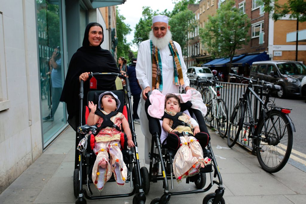
Rare conjoined twins, Safa and Marwa, have been separated at a hospital in London. The 19-month-old girls were craniopagus twins, with their skulls and blood vessels fused together. This made the procedure extremely complicated and it took three major operations before the girls could be discharged. The operations added up to more than 50 hours of surgery time and involved 100 members of staff from Great Ormond Street.
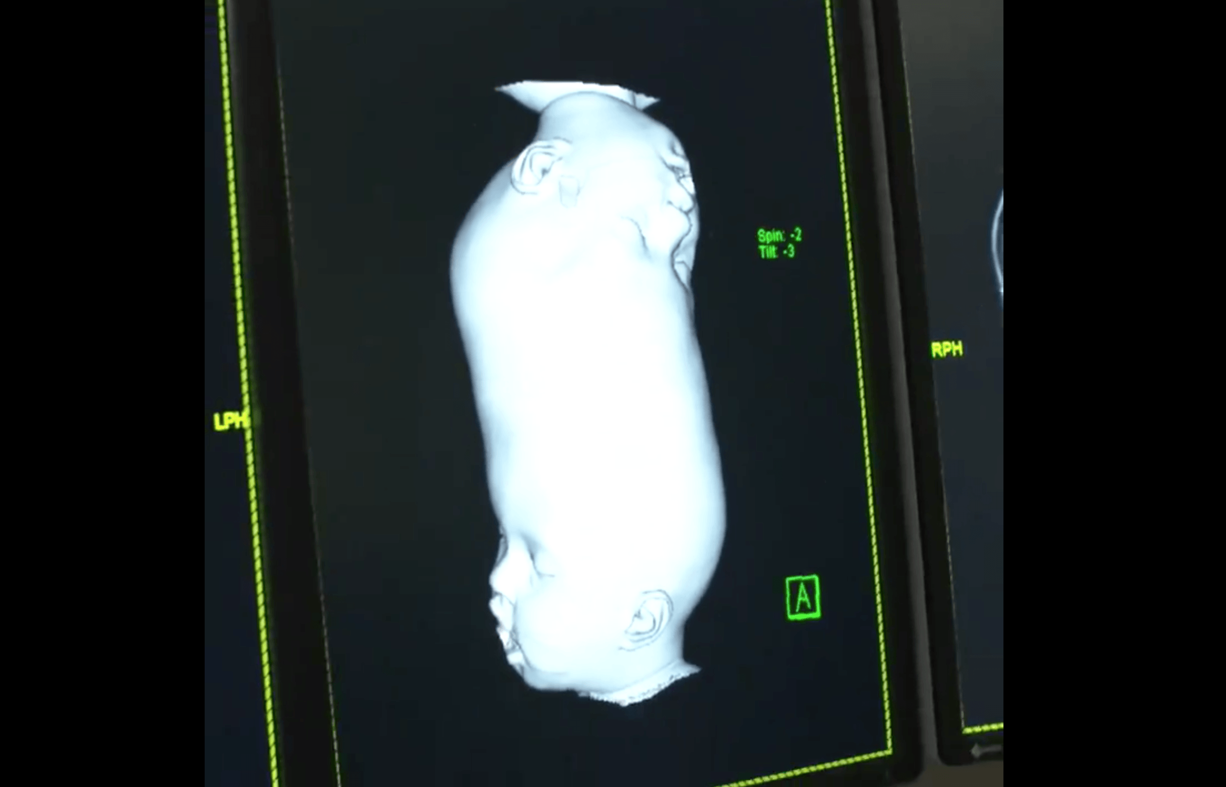
Great Ormond Street Hospital has previously successfully separated craniopagus twins in 2006 and 2011. Safa and Marwa’s separation earlier this year was aided by state-of-the-art technology not available during previous operations. For the first time, the team used virtual reality, advanced imaging and 3D anatomical printing to prepare for the operation.
Thanks to the new technology, the team used images of the girls’ brains and blood vessels to plan and practice the surgery in advance to minimise complications. VR was used to exact a replica of the twins’ anatomies. This helped the doctors to practise the surgery before the first cut was made and visualise the complexities of the girls’ skull as well as the position of their brains and blood vessels.
Professor Dunaway, head of the hospital’s craniofacial unit, said in a statement: “From the surgical teams, scientists and engineers who helped us plan and perform the operations to the paediatricians, nursing staff, physiotherapists and occupational therapists, it has been a huge team effort and every single person has played a key part in helping Safa and Marwa.”